
This web page was produced as an assignment for an undergraduate course at Davidson College.
Researchers reveal changes in proteins, mRNA, non-essential amino acids, and genes during grizzly bear hibernation that contribute to atrophy resistance
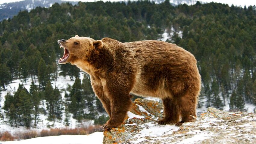
Grizzly bears hibernate for 5-7 months without losing muscle, but if other organisms such as humans were immobilized or restricted calories for that duration their muscles would waste away. Although it is already known that grizzly bears utilize fat reserves and reabsorb urea for maintaining blood glucose levels and avoiding using amino-acids from muscle proteins, the contribution of muscle cells in the resistance to muscle atrophy is unknown (Nelson 1973; Giroud et al. 2019). Mugahid and his colleagues answered this question using the skeletal muscle proteome and transcriptome of grizzly bears revealing the molecular changes that limit atrophy and evaluated the evolutionary conservation of these mechanisms. Through a series of experiments, they concluded that many proteins regulated during hibernation are involved in the activity of the TCA cycle and Pi3k-Akt pathway, non-essential amino acids are increased in hibernation and can be supplemented to decrease atrophy, different genes are regulated in hibernation and are evolutionarily conserved, and Pdk4 and Serpinf1 genes acquired later in evolution contribute to atrophy.
First, they performed mass spectrometry on all the proteins from muscle biopsies of cubs and bears before and during hibernation which revealed 66 proteins that are regulated during hibernation, many of which are involved in metabolic pathways such as the TCA cycle, a process that releases stored energy. Sequencing the RNA of these biopsies revealed more protein transcripts that are associated with the Pi3k-Akt pathway which regulates organ growth and metabolism (Stitt et al. 2004). During hibernation, activity of this pathway increases. For example, they found that the insulin-sensitive receptor substrate Irs-1 is upregulated along with its downstream effector and upstream binding proteins, and the less sensitive insulin receptor Irs-224 is suppressed.
Next, the researchers compared non-essential amino acids in muscles of hibernating bears and aging humans because they affect the muscle’s ability to maintain protein levels. Non-essential does not mean that organisms don’t need them, but they are amino acids the body produces instead of receiving from food. Of the 11 non-essential amino acids, 6 increased in hibernation and 7 decreased in aging humans. Due to this finding, the researchers wanted to know how supplementing non-essential amino acids would affect muscle atrophy. To evaluate this, they treated muscle cells with synthetic glucocorticoid dexamethasone (Dex) to model atrophy or a vehicle as a control. After supplementing both groups with a 10 fold elevated level of non-essential amino acids, levels of MT-1 and Ube2b mRNA decreased, but the other two atrophy markers were not affected. Along with decreased atrophy markers, amino acid treated muscle cells were also larger. The researchers attributed this change is muscle size to increased phosphorylation of S6k which they also reported in the amino acid treated group and in hibernating bear muscle.
Their next goal was to identify previously unknown target genes that regulate muscle mass. Using public atrophy-related data sets of humans and mice and their collected data from hibernating bears, they identified 18 genes regulated specific to hibernation, 6 genes regulated in non-hibernation samples, and 2 genes regulated in both. Algorithms revealed that the genes regulated in non-hibernation samples are often co-expressed and therefore are part of similar functional groups.
To understand if these genes were evolutionary conserved, they knocked down homologous genes in C. elegans and determined their effects on worm size and speed. Only 4 genes affected worm perimeter, and 9 affected their speed. Therefore, the researchers concluded that these genes are evolutionarily conserved, but there is stronger evolutionary pressure on those that pertain to muscle function.
They next questioned if the genes not validated in C. elegans were acquired later in evolution by examining two genes with roles in muscle related signaling pathways: Pdk4, which results in protein degradation, and Serpinf1, which can be linked to muscle wasting (Lee et al. 2015; Cai et al. 2004). Muscle cells were first treated with dexamethasone to model atrophy or a vehicle as a control like before. Then the researchers decreased the expression of Pdk4, and quantified the expression of the same atrophy markers from earlier. They found that MAFbx mRNA levels increased and Murf1 levels decreased in both experimental and control models when Pdk4 expression was decreased, but Ube2b and MT1 levels didn’t change. When they decreased Serpinf1 expression next, the expression levels of all four markers decreased suggesting that Serpinf1 affects atrophy signaling more. Lastly, they examined the effects of Pdk4 and Serpinf1 on muscle size by creating knockdowns again. Knockdowns of both genes resulted in larger muscles compared to controls, but Serpinf1 knockdowns had the largest muscle sizes.
Overall, this proteomic and transcriptomic analysis of hibernating grizzly bears revealed the proteins, pathways, non-essential amino acids, and genes that contribute to atrophy resistance. Most notably, it seems that increased non-essential amino acids plays one of the largest roles in preserving muscle mass. This study also presents decreased Pdk4 expression as promising for muscle cell size regulation, but Pdk4 protein is typically increased during hibernation (Buck et al. 2002). Therefore, these findings should be assessed by other researchers to evaluate this controversy. This study also leads to future research into potential muscle atrophy therapies such as using non-essential amino acids to suppress atrophy signaling. Such studies could aid in patient recovery from muscle diseases and benefit immobilized people who cannot maintain muscle mass.
Sommer Holmes is currently enrolled in Davidson College. Contact her at soholmes.davidson.edu.
References
Buck, M. J., Squire, T. L. & Andrews, M. T. Coordinate expression of the PDK4 gene: a means of regulating fuel selection in a hibernating mammal. Physiol. Genomics 8, 5–13 (2002).
Cai, D. et al. IKKbeta/NF-kappaB activation causes severe muscle wasting in mice. Cell 119, 285–298 (2004).
Giroud, S. et al. Lipidomics Reveals Seasonal Shifts in a Large-Bodied Hibernator, the Brown Bear. Front Physiol 10, 389 (2019).
Lee, S. J. et al. Pyruvate Dehydrogenase Kinase 4 Promotes Vascular Calcification via SMAD1/5/8 Phosphorylation. Sci Rep 5, 16577 (2015).
Mugahid, D.A., Sengul, T.G., You, X. et al. Proteomic and Transcriptomic Changes in Hibernating Grizzly Bears Reveal Metabolic and Signaling Pathways that Protect against Muscle Atrophy. Sci Rep 9, 19976 (2019).
Nelson, R. A. Winter sleep in the black bear. A physiologic and metabolic marvel. Mayo Clin. Proc. 48, 733–737 (1973).
Stitt, T. N. et al. The IGF-1/PI3K/Akt Pathway Prevents Expression of Muscle Atrophy-Induced Ubiquitin Ligases by Inhibiting FOXO Transcription Factors. Mol. Cell 14, 395–403 (2004).
Return to homepage.
© Copyright 2020 Department of Biology, Davidson College, Davidson, NC 28036.