This web page was produced as an assignment for an undergraduate course at Davidson College.
A transcriptome and proteome approach reveals differential regulation that gives context to the regeneration of gecko tails after amputation.
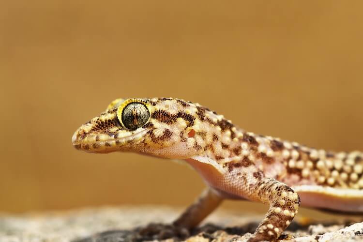
Epimorphic regeneration is a phenomenon that involves extensive cell proliferation of somatic stem cells, differentiation, reformation, as well as blastema formation (Yokoyama, 2008). Blastemas, which are masses of cells capable of growth and regeneration into organs or body parts, play a key role in epimorphosis resulting in animal limb regeneration (Kragl et al., 2009). True epimorphic regeneration is rarely observed in mammals, but the ability to regenerate full limbs has been studied in certain amphibians, fishes, and lizards (Londono et al., 2018). This study specifically focuses on epimorphic regeneration in Hemidactylus frenatus, the house gecko. As a result of their flight response from predators, lizards have the ability to self-detach and amputate their tails. After this detachment, multiple cellular responses are activated to begin the course of regeneration. Many genes or proteins have been shown to influence epimorphic regeneration, but the underlying mechanisms of relevant molecules remain unknown.
This study sought to identify the molecular mechanisms of the tail regeneration process during its early stages and to determine which molecules were involved in jump starting the regenerative processes in geckos. Researchers used transcriptome and proteome approaches to list out crucial genes and proteins that are differentially expressed, and what the expression means for the molecular processes of regeneration.
First, researchers analyzed the transcript sequences from gecko tail tissue samples. Researchers found a total of 417 genes that were associated with regeneration of gecko tail tissue. A total 254 genes were further analyzed for expression patterns at different points in the regeneration process. Expression of genes was analyzed at 1, 2, and 5 days post-amputation. 26, 38 and 29% of identified genes corresponding to the days post-amputation were found to be upregulated. With this, 54, 35 and 46% of genes were found to be downregulated at the three time marks. All of the genes appeared to be highly enriched for a certain functional the early developmental process for some function. Observed was significant changes in genes associated with anatomical structure development and molecular functions. The specifics of these genes highlighted a crucial involvement of genes that initiated wound healing. Major genes associated with growth, cell proliferation, immune response activation, and ion binding were found to be involved in the onset of the healing phase. Gene analysis also showed groups of genes that were upregulated at 1 day post-amputation, but downregulated at 2 and 5 days post-amputation and vice versa. Analysis showed that gene expression at 2 and 5 days was clustered with each other and more prominent than the expression patterns at 1 day.
Next, researchers analyzed the involvement of the proteome in the early stages of gecko tail regeneration. A total of 128 proteins were found to be differentially regulated during the observed stages of generation. 36 and 33% of proteins were seen to be upregulated at 1 and 2 days, while only 10% were found to be downregulated. 67% of proteins were found to be downregulated for the regeneration mechanism at 5 days with only one protein being upregulated at that point. A total of 37 proteins that were differentially regulated at the proteome level were also found to be differentially regulated at the gene level based on the transcriptome analysis. Like the transcriptome expression patterns, the protein expression patterns were also found to be associated with clustering at days 2 and 5. All proteins that were upregulated at 1 day post- amputation were downregulated at day 2 or 5. All downregulated proteins at day 1 were upregulated at day 2 or 5. Researchers were able to see distinct patterns of upregulation and downregulation of the transcriptome and the proteome, but they also want to understand what these patterns actually entail for gecko tail regeneration. Based on the aforementioned analyses, it was seen that 29, 26, and 20% of genes/proteins were found to be upregulated for the initiation of regeneration mechanisms. Related to this, 39, 24 and 53% of genes/proteins were found to be downregulated for regeneration mechanisms. A multitude of crucial genes/proteins that are associated with pain and inflammation, DNA repair, proteasome activities, and cellular morphology were upregulated at 1 day post-amputation. Subsequently, downreuglation of these genes and proteins related to inflammation was observed at 2 days post-amputation. Also on day 2, genes and proteins that related to skeletal and muscle development processes were observed to be upregulated. With this, on day 5 post-amputation, all genes and proteins involved in inflammation and other post-translational modifications were found to be downregulated, even as they were upregulated at days 1 and 2.
As previously mentioned, the main goal of this study was to gain a greater insight into the genomic and proteomic changes of tail tissue regeneration in the house gecko. The differential regulation and expression of genes and proteins identified numerous cellular processes and canonical pathways that are turned on or off during different stages of epimorphic regeneration. This study helps to corroborate the data observed in similar studies that looked at regeneration in zebrafish and other gecko species. With this data, we can also start to get more of an understanding of how similar genes and proteins are regulated in humans. This greater understanding could lead to new advancement in how human wounds are repaired, and how we can work in conjunction with our transcriptomes and proteomes to promote tissue regeneration. This study is just a piece of the puzzle that is epimorphic regeneration.
Miles Davis is a junior biology major at Davidson College. Contact him at: midavis@davidson.edu
© Copyright 2020 Department of Biology, Davidson College, Davidson, NC 28036.