This web page was produced as an assignment for an undergraduate course at Davidson College.
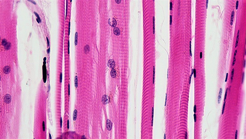
Analysis of the proteome in muscle fibers showcased differential adaptations, revealing mechanisms important to muscle function and health.
.jpg)
Over the past few decades, the importance of exercise and healthy eating has been stressed through all types of media (i.e. documentaries, ad campaigns, school curriculum, etc.). Additionally, medical professionals have continued to recommend consistent exercise as one method to mitigate and prevent diseases. The study by Deshmukh et al. continues on the information provided by these social changes to provide insight on how the regulatory processes, specifically within the muscle, change as a result of endurance training over 12 weeks. However, muscle is made up of fibers. These fibers are categorized into two types: slow-twitch and fast-twitch. Slow-twitch is more resistant to fatigue and highly expresses oxidative enzymes that are important to the mitochondrial production of ATP (Deshmukh et al. 2021). Fast-twitch fibers are further categorized into two types, type IIa and IIx, and they rely on enzymes involved in glycolysis to rapidly produce energy for them to contract (Deshmukh et al. 2021. While slow- and fast-twitch fibers are involved in different metabolic processes, their different roles in the body offer a unique study opportunity on their individual adaptations apart from one another. Deshmukh et al. observed these properties and instead of focusing on the overall quadriceps muscle, they focused on its individual fibers. The fiber-specific metabolic processes and regulatory pathways offer potentially differing changes throughout a muscle group in response to endurance training, and thus, can provide novel therapies targeting their regulatory pathways.
This study wanted to analyze muscle changes which required taking samples from participants and conserving those samples so that their fibers could be isolated for analysis. The biopsy samples were taken from five men before and after 12 weeks of endurance training (specified as 1 hour of cycling at 75-90% of the subject’s max heart rate). Samples were then instantly frozen and freeze-dried. Then, fibers were individually separated from the muscle samples to perform proteomic analysis, Deshmukh et al. utilized sequential digestion and high-resolution mass spectrometry (MS). Sequential digestion is a technique that involves lysis which breaks down cell membranes to release the cell’s protein contents for analysis(Peng and Gygi 2001). In this study, lysis was performed on single fibers to observe the major and minor differences between slow- and fast-twitch fibers. Sequential digestion then utilizes antibodies to isolate proteins from cell contents in order to quantify them in MS. However, before the proteins are run through MS, their peptides are broken down by enzymes for easier quantification by the technology. This study utilized the enzymes Trypsin and LysC. MS then quantifies how many proteins are within the mixture and allows for their identification (Aebersold and Mann 2003).
Within this study, they were able to identify 4,158 proteins from both types of fibers using MS. The next step was to understand how the regulation of these proteins changed due to exercise and if those changes are dependent on the fiber type. Ultimately, Deshmukh et al. separated proteins quantified by MS in only one of the two fiber types and labeled them “exclusive”. Using that method, they found that 131 proteins were regulated differentially depending on fiber type. In slow-twitch fibers, 61 proteins were significantly regulated by exercise training and 13 proteins in fast-twitch. Deshmukh et al. wanted to understand the functional role the 61 proteins in slow-twitch and the 13 proteins in fast-twitch had in the cell. However, due to these proteins belonging to a vast range of biological processes, there was no clear functional identification. Future studies that can validate these proteins’ roles in a detailed fashion may be able to understand muscle metabolism on a fiber-type level. Although the roles of these proteins could not be specified and validated, the study concluded that the metabolic processes that these twitch fibers rely on may have a role in how regulatory processes adapt to exercise. One observation of a dramatic decrease in glucose-6 phosphate-dehydrogenase (G6PD), an enzyme that works in many metabolic pathways and negatively regulates muscle insulin sensitivity (Deshmukh 2021), showed that exercise had a positive affected by increases in insulin sensitivity and glucose uptake in slow-twitch fibers.
Overall, this study was able to identify and quantify fiber-type-specific proteomes and assess how they are regulated by exercise, providing information on specific adaptations to metabolic processes. Understanding the effects exercise has on intracellular signaling has the ability to change the way we use medicines and therapies in general especially in cases where there are diseases that directly affect metabolic processes (Vina et al. 2012). As exercise tends to become habitual and a part of a routine, this study affirms the work of many medical professionals to encourage diet and exercise before turning over to drug therapies. However, this study only tested the effects on muscle fibers from one type of exercise on a subject pool of five men. To really consider the possibilities, further studies should expand the subject pool to women, diversify age ranges, and explore exercise intensity, type, and duration. The more information on how exercise provides the body with better metabolic regulation, the more likely it will be that exercise plans can assist as a metabolic therapy or for other regulatory pathways. As diagnoses of childhood obesity and metabolic diseases like type II diabetes rise across the United States, exercise can provide one method for metabolic regulation without the use of pharmaceutical medicines (Hawley et al. 2014).
Kamryn Graham is a sophomore biology major at Davidson College. Contact her at kagraham@davidson.edu.
References
Aebersold, R. & Mann, M. Mass spectrometry-based proteomics. Nature422, 198–207 (2003).
Deshmukh, A. S. et al. Deep muscle-proteomic analysis of freeze-dried human muscle biopsies reveals fiber type-specific adaptations to exercise training. Nature Communications12, 304 (2021).
Hawley, J. A., Hargreaves, M., Joyner, M. J. & Zierath, J. R. Integrative Biology of Exercise. Cell159, 738–749 (2014).
Peng, J. & Gygi, S. P. Proteomics: The Move to Mixtures. Journal of Mass Spectrometry36, 1083–1091 (2001).
Vina, J., Sanchis‐Gomar, F., Martinez‐Bello, V. & Gomez‐Cabrera, M. C. Exercise acts as a drug; the pharmacological benefits of exercise. British Journal of Pharmacology167, 1–12 (2012).